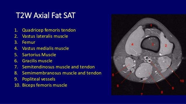
Knee Muscle Anatomy Mri - Mri anatomy of knee Dr. Muhammad Bin Zulfiqar / The knee joint is the junction of the thigh and leg.. Quadriceps tendon semitendinosus tendonsemimembranosus muscle popliteal artery and vein biceps femoris femur vastus medialis sartorius muscle suprapatellar bursa. Injuries of the patellofemoral joint. An understanding of normal anatomy and biomechanics of the knee extensor mechanism is necessary to comprehend the imaging of extensor mechanism injuries. Magnetic resonance imaging (mri scan): Normal anatomy, variants and checklist.
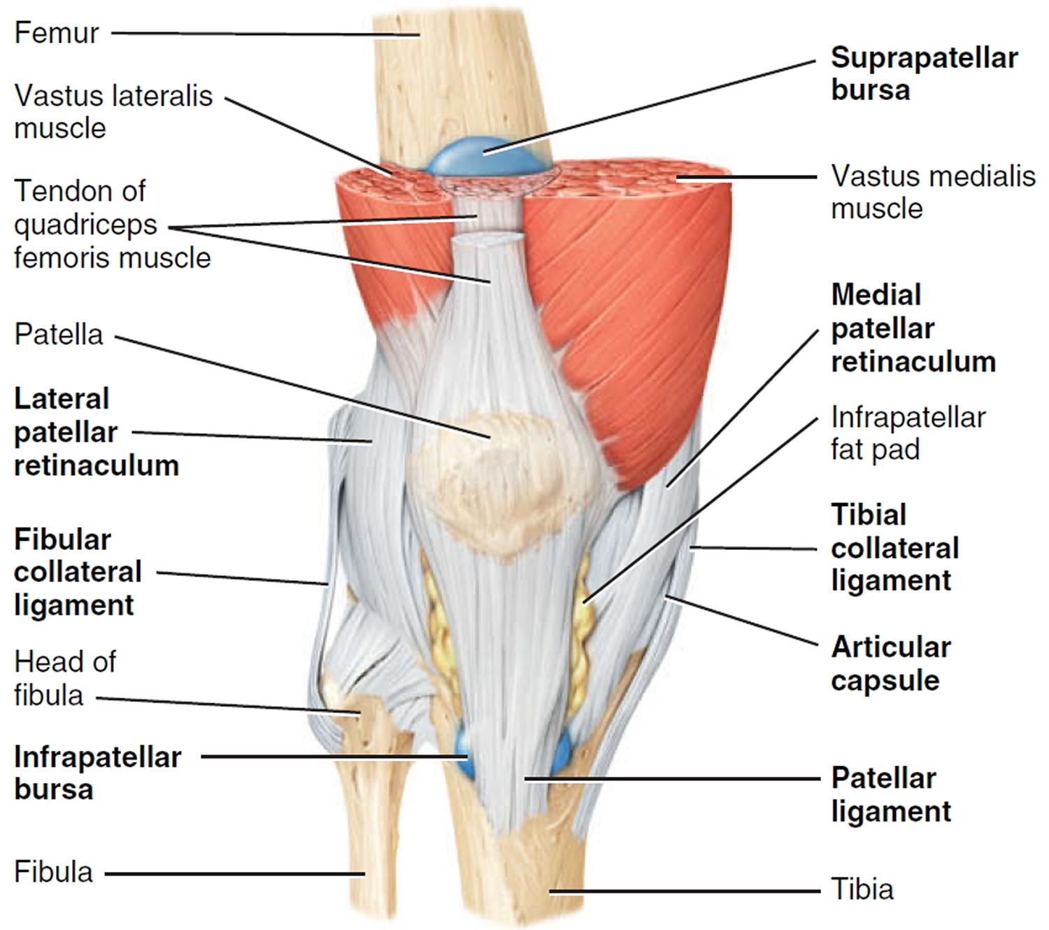
Each anatomical structure was labeled interactively. Use the checklist to quiz yourself. This mri knee cross sectional anatomy tool is absolutely free to use. They are attached to the femur (thighbone), tibia (shinbone), and fibula (calf bone) by fibrous tissues called ligaments. Please email baodo at stanford.edu.
Mri anatomy of knee Dr. Muhammad Bin Zulfiqar from image.slidesharecdn.com 4, infrapatellar fat pad of hoffa. Free access interactive and dynamic anatomical atlas. There are various muscles that control movement, ligaments that. This section of the website will explain. Tendons attach the muscles to each other. Robin smithuis and henk jan van der woude. Current and accurate information for patients about magnetic resonance imaging (mri)of the knee. Magnetic resonance imaging (mri scan):
Please email baodo at stanford.edu.
This mri knee cross sectional anatomy tool is absolutely free to use. The muscles of the knee include the quadriceps, hamstrings, and the muscles of the calf. Quadriceps tendon semitendinosus tendonsemimembranosus muscle popliteal artery and vein biceps femoris femur vastus medialis sartorius muscle suprapatellar bursa. 4, infrapatellar fat pad of hoffa. Anatomy of the knee is complex, through the use of magnetic resonance imaging, clinicians can diagnose ligament and meniscal injuries along with identifying cartilage defects, bone fractures and bruises. Find out how the different structures fit together in our knee diagram the knee joint is the largest and one of the most complex joints in the human body. The knee joint is the junction of the thigh and leg. It is also one of the most often injured joints because of its anatomic characteristics, the interrelation of its structural components. Mri for evaluating knee pain in older patients: They are attached to the femur (thighbone), tibia (shinbone), and fibula (calf bone) by fibrous tissues called ligaments. Anterior graphic of the shoulder. Learn about the muscles, tendons, bones, and ligaments that comprise the knee joint anatomy. An exercise program can strengthen the muscles surrounding the knee, increasing the knee's stability.
4, infrapatellar fat pad of hoffa. Each anatomical structure was labeled interactively. Use the checklist to quiz yourself. Injuries of the patellofemoral joint. Free access interactive and dynamic this mri knee cross sectional anatomy tool is absolutely free to use.
Knee Pain - Causes, Exercises, Remedies, Medication ... from healthjade.com Find out how the different structures fit together in our knee diagram the knee joint is the largest and one of the most complex joints in the human body. Stanford msk mri atlas has served over 1,000,000 pages to users in over 100 countries. They are attached to the femur (thighbone), tibia (shinbone), and fibula (calf bone) by fibrous tissues called ligaments. The muscles that affect the knee's movement run along the thigh and calf. Involved early gray = muscle: Technical considerations for mri evaluation of the knee extensor mechanism. There are various muscles that control movement, ligaments that. This section of the website will explain.
Each anatomical structure was labeled interactively.
The knee joint is the junction of the thigh and leg. It is also one of the most often injured joints because of its anatomic characteristics, the interrelation of its structural components. Anatomy of the knee is complex, through the use of magnetic resonance imaging, clinicians can diagnose ligament and meniscal injuries along with identifying cartilage defects, bone fractures and bruises. Free access interactive and dynamic anatomical atlas. Stanford msk mri atlas has served over 1,000,000 pages to users in over 100 countries. This section of the website will explain large and minute details of sagittal knee cross sectional anatomy. Magnetic resonance imaging (mri scan): Scroll through the structures to understand the anatomy. There are various muscles that control movement, ligaments that. Normal anatomy, variants and checklist. How does the knee joint work? Mri for evaluating knee pain in older patients: This webpage presents the anatomical structures found on knee mri.
Which are the ligaments that keep it stable? 4, infrapatellar fat pad of hoffa. Anterior graphic of the shoulder. Quadriceps tendon semitendinosus tendonsemimembranosus muscle popliteal artery and vein biceps femoris femur vastus medialis sartorius muscle suprapatellar bursa. Normal anatomy, variants and checklist.
knee anatomy mri - DriverLayer Search Engine from konez.com Mri uses a powerful magnetic field, radio waves and a computer to produce detailed. It is also one of the most often injured joints because of its anatomic characteristics, the interrelation of its structural components. Which are the ligaments that keep it stable? Learn about the muscles, tendons, bones, and ligaments that comprise the knee joint anatomy. How does the knee joint work? Please email baodo at stanford.edu. The muscles of the knee include the quadriceps, hamstrings, and the muscles of the calf. Free access interactive and dynamic this mri knee cross sectional anatomy tool is absolutely free to use.
4, infrapatellar fat pad of hoffa.
This mri knee cross sectional anatomy tool is absolutely free to use. The journal of musculoskeletal medicine. Click on the links to show each structure. This section of the website will explain. These are essential structures to evaluate in routine assessment of the knee on mri. This mri knee cross sectional anatomy tool is absolutely free to use. Learn anatomy using a full pacs! Use the checklist to quiz yourself. Scroll through the structures to understand the anatomy. Please email baodo at stanford.edu. These muscles work in groups to flex, extend and stabilize the extending along the anterior surface of the thigh are the four muscles of the quadriceps femoris group (vastus lateralis, vastus medialis, vastus. Robin smithuis and henk jan van der woude. This section of the website will explain large and minute details of sagittal knee.


0 Comments:
Posting Komentar